UHN Research & Innovation Cores Newsletter
Dear Readers,
Welcome to the August 2021 issue of the UHN Research & Innovation Cores Newsletter!
We have had a great response to our last edition and want to thank all of you who have taken the time to read it.
This edition takes an in-depth look at the Flow Cytometry facilities providing access to several state-of-the-art analytical flow cytometers, high-speed cell sorting, and mass cytometry services.
Also featured are The Princess Margaret Living Biobank (PMLB), which offers Patient-Derived tumour Organoid (PDO) and Xenograft (PDX) models and associated services for research studies as well as the The Advanced Optical Microscopy Facility (AOMF), which provides optical microscopy technologies for the observation of fixed cells, live cells, and tissues.
Thanks for reading our newsletter!
Luke Brzozowski
UHN Research & Innovation Cores
Flow Cytometry
Flow cytometry is a technology that can be used to detect, count, sort and profile cells that have been fluorescently labelled. This technology can be used to evaluate cells from blood, body fluids or tumours and, as such, is integral to many research programs.
UHN has three Flow Cytometry core facilities, the Princess Margaret (PM) Flow Cytometry Core, the Krembil Discovery Tower (KDT) Flow Cytometry Core and the Princess Margaret Cancer Research Tower (PMCRT) Flow Cytometry Core. These facilities are available to support UHN researchers as well as the broader research community.
Recent investments have been made at all three facilities to provide access to state-of-the-art techniques and instruments supported by a team of experts that provide consultation, technical support, and training.
The PM Flow Cytometry Core has recently introduced Spectral Flow Cytometry technology with a new Sony ID7000 Spectral Cell Analyzer. These Analyzers use a technique wherein a spectrograph and multichannel detector replace the traditional mirrors, optical filter, and photomultiplier tubes. This method broadens a fluorophore’s spectral profile by capturing the entire visible and near-IR spectrum.
The KDT Flow Cytometry Core will be joining the PM Core by implementing Stratocore, an innovative cloud-based software that researchers at KDT and anywhere across UHN can use to reserve equipment. As the go-live date approaches, the KDT Flow Cytometry Core will notify current KDT users to ensure they take advantage of this operational improvement.
The PMCRT Flow Cytometry Core will be offering researchers access to new sorters that will provide precise sorting capabilities and new analyzers that will enable more sophisticated multi-colour analysis. These new instruments are compatible with other existing platforms to allow for a seamless transition from cell isolation to cell analysis.
The Princess Margaret Living Biobank
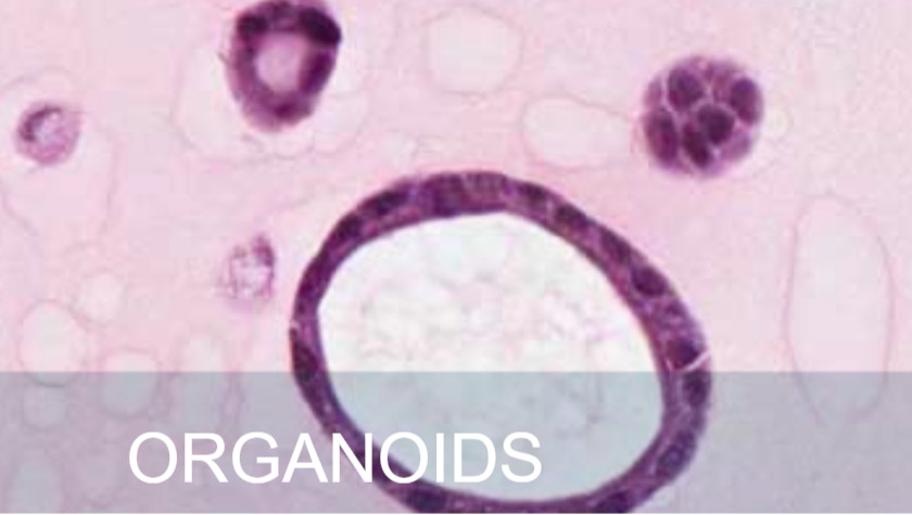
Patient-derived xenograft (PDX) models and patient-derived organoids (PDO) are important tools in translational oncology research. Implanting patient tumour tissue into mice or a gel matrix, respectively, creates a clinically relevant system to study cancer, test the efficacy of cancer therapies, and develop personalized medicine approaches.
The Princess Margaret Living Biobank (PMLB) has extensive patient-derived tumour models, including but not limited to ovarian, head and neck, lung, breast, pancreas, and colon cancers. These models are available for use by UHN teams and other external academic or private-sector researchers.
As a core facility, PMBL acts as a repository for REB-approved and patient-consented models, which includes associated multi-omics and clinic-pathological data. These models are annotated by molecular and patient-matched clinic-pathological data. PMBL is a resource for annotated patient-derived tumour models and their derivatives (i.e.; formalin-fixed, frozen, and fresh specimens).
This facility offers many services to researchers, including model establishment, drug screening, functional screening, and biomarker discovery. Additionally, PMBL provides training and offers consultation based on extensive expertise on organoid and xenograft studies.
The Advanced Optical Microscopy Facility
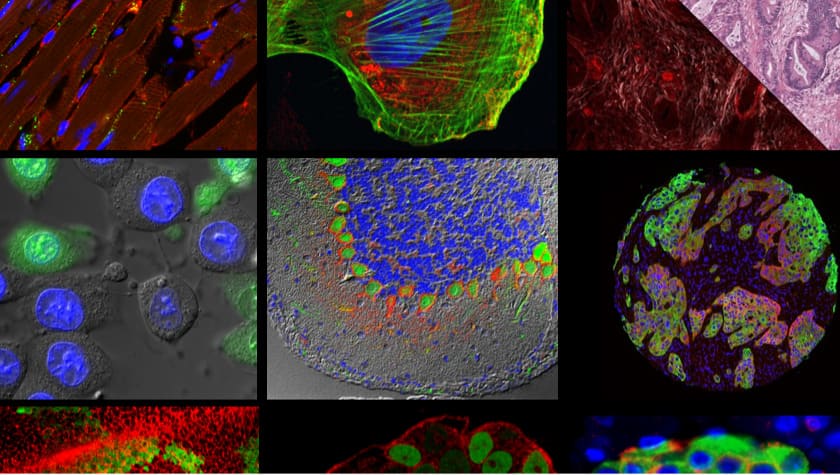
The Advanced Optical Microscopy Facility (AOMF) is the largest core facility of its type in Canada. Through collaboration and integration of microscopy laboratories across UHN, AOMF has become a sustainable core facility that supports over 600 researchers a year.
With over 40 instruments at four UHN sites, AOMF offers UHN researchers and the broader researcher community access to a wide array of microscopy technologies and the expertise of the AOMF team.
AOMF has a range of cutting-edge microscopes and imaging systems, including confocal, multiphoton, widefield, super-resolution, tissue slide scanners and more. While confocal microscopes are the most popular instruments, AOMF is home to several specialty instruments such as super-resolution (STED, GSD), Raman, and Total Internal Reflection Fluorescence (TIRF) microscopes.
AOMF also provides full-service brightfield and fluorescence slide scanning, digitizing entire slides in high resolution. These images are used in tandem with very powerful pattern-recognition software, which provides cell by cell quantification of entire tissue sections.
Staff at AOMF come from diverse backgrounds in mathematics, physics, chemistry, and biology and have many years of experience in the field of microscopy. This creative and innovative team provides full-service slide scanning, image analysis, microscopy and image analysis training to researchers, students, and industry partners. The AOMF core facility also offers comprehensive courses and tailored workshops on microscopy to trainees from across the country.
If you have a sample that fluoresces or interacts with light in some way, AOMF can image it or extract information from it to support your research.