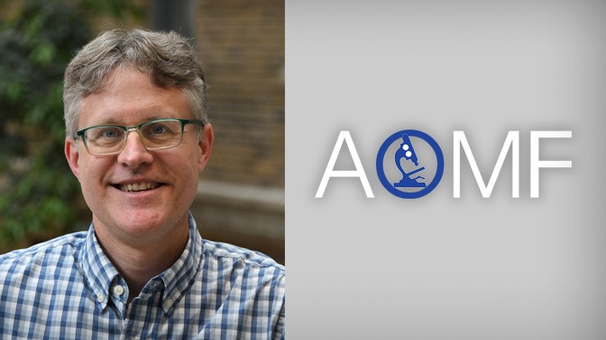
An Interview with James Jonkman

An interview with James Jonkman, Staff Scientist & Manager of the AOMF facility at UHN
Tell us about your professional background.
During my graduate studies at the University of Waterloo my fascination with lasers, optics and confocal microscopy sparked my passion for the field of optical microscopy. This drove me to develop a training program at the Princess Margaret and later led me to the European Molecular Biology Laboratory (EMBL) in Germany. While there, I worked in Ernst Stelzer's lab, which excels in the field of optical microscopy. In 2002, back in Toronto, I proposed the transformation of a single confocal microscope into a core facility, giving birth to the AOMF. Since then, I have expanded the facility, integrated other microscopy sites, and continued to acquire advanced imaging equipment. The AOMF is presently the largest optical microscopy facility in Canada.
What are your responsibilities at AOMF and what aspect of your role do you enjoy the most?
As the manager of the AOMF, an important part of my role is to stay abreast of the latest microscopy advancements. I do this by attending conferences, webinars and instrument demos. I consult with clients, troubleshoot instrumentation issues and provide training sessions. I also help guide new acquisitions and write descriptions and justifications for grant applications.
One of the most rewarding aspects of my job is teaching and training users, through which I strive to promote scientific rigor and reproducibility. Together with my staff, I have developed comprehensive training materials for each instrument under our care, offering personalized, hands-on sessions and a Fundamentals of Microscopy course. I actively engage with the microscopy community, through organizing webinars and leading the Optical Microscopy Users Group (O-MUG) meetings. Over the years, I have co-authored book chapters, review articles and tutorial papers. These publications, including a Nature Protocols tutorial on quantitative confocal microscopy, showcase my dedication to teaching users how to obtain meaningful data from their experiments.
How does the AOMF help the research community advance their research?
We offer much more than just confocal microscopes. Our services include brightfield and fluorescence whole-slide scanning, combined with advanced HALO analysis software. We offer exclusive technologies, such as resonant-scanning confocals, multiphoton microscopes, and super-resolution microscopes, we can capture dynamic processes and image deep into tissues with exceptional resolution. Our capabilities extend to live-cell imaging, clearing techniques, organoids, nanoparticles, microfluidic devices and more. We provide personalized tours and consultations to determine the best microscope or technique for each user. Our knowledgeable staff can also train users on advanced analysis software to transform their images into valuable data.
Collaboration is essential for good science. How does the AOMF promote teamwork?
Collaboration lies at the heart of scientific progress, and AOMF actively fosters collaboration among cores and principal investigators. I have been active in the microscopy community for years. During my tenure as a co-president and vice-president of the Canadian Cytometry and Microscopy Association (CCMA), I organized national symposia, launched a newsletter and led the website revamp. I also played a key role in establishing the Canadian Network of Scientific Platforms (CNSP) and currently serve on the Executive Board of Canada Bioimaging. As a member of various international associations, such as the Association of Biomolecular Resource Facilities (ABRF) and Bio-Imaging North America (BINA), I am continuously learning from imaging core facilities worldwide. This perspective allows me to bring best practices to the AOMF and Wright Cell Imaging Facility (WCIF).
What is next for the AOMF?
Across our four UHN sites, we are always ready to take on new challenges and we bring enthusiasm to every project that we work on with UHN researchers and the wider community. As we look ahead, we aim to enhance our roster in order to meet growing demand. We have plans to acquire a second confocal microscope for our Krembil site to address overbooking issues. We are replacing end-of-life systems at our Toronto General Hospital site and adding a new confocal. We are also currently securing funding for a light sheet microscope at our Krembil site to eliminate the need for external resources.
At the AOMF, I have been lucky to be able to advance microscopy in Canada and to help foster a community of individuals that are passionate about imaging. As we progress, I am excited to witness the remarkable biomedical discoveries and breakthroughs enabled by the AOMF.
For more information on AOMF, please visit: www.aomf.ca